

Computed Tomography (CT)
A Computed Tomography (CT) exam is a medical imaging test that helps Radiologists see detailed pictures of the inside of your body. It uses X-rays and computer technology to create cross-sectional images of organs, bones and blood vessels.
What to Expect
On Arrival
You’ll be greeted by one of our friendly technologists.
You will be guided to a changing room and be asked to change into a dressing gown. You will also be asked to remove metal items such as jewelry and body piercings.
The Technologist will then guide you to the CT room and position you on the CT table for your exam.
During the Procedure
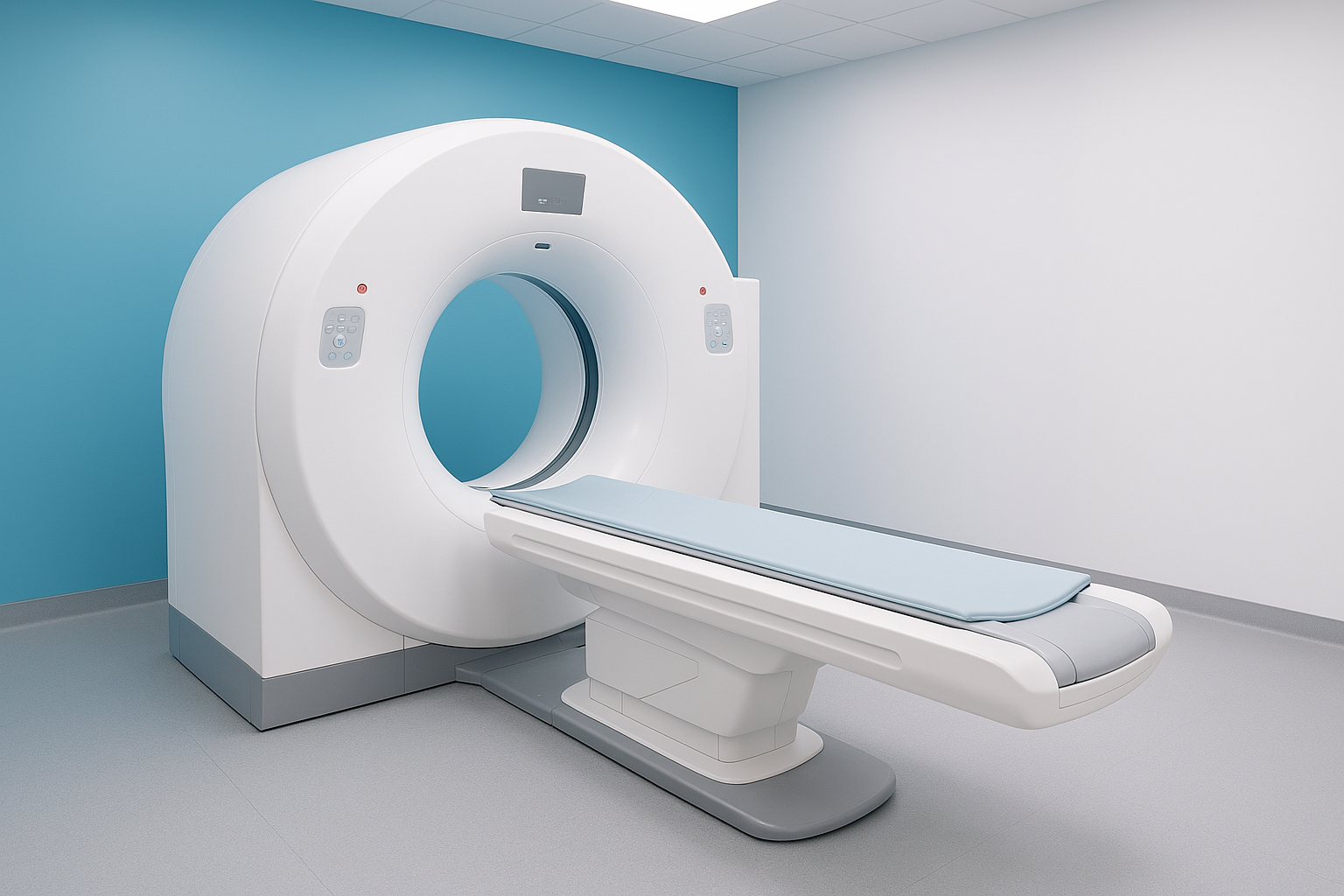
You will lie on a padded table that moves through a short, donut-shaped scanner. The scanner never “closes” around you. We provide warm blankets and help with positioning and comfort.
If needed for certain exams you will be asked to hold your breath for a brief moment during the scan.
You’ll be able to hear and talk to the technologist while the scan is in progress.
After the Procedure
You can return to normal activities immediately.
Your doctor will receive results within 1-2 business days.